Studying adipogenesis and adipocyte biology requires the isolation of primary preadipocytes from adipose tissues. However, primary preadipocytes have a limited lifespan, can only undergo a finite number of divisions, and often lose their original biological characteristics before becoming senescent. The repeated isolation of fresh preadipocytes, particularly from young pups or agedanimals, is costly and time consuming. Immortalization of these cells offers asolution by overcoming cellular senescence and maintaining proliferative capacity, allowing for long-term studies without the continuous need to isolate new cells from animals. Immortalized cell lines thus provide a consistent and reproducible experimental model, significantly reducing variability across different animals.
To better understand and investigate the roles of brown and white adipocytes in energy homeostasis, thermogenesis, metabolic disorders, and aging processes in vitro, we established this immortalized mouse brown preadipocyte cell lines from old mice(12-month-old and 27-month-old). One group of old mice (12 months) was also maintained on a normal diet and a high-fat, high-fructose diet for 10 months.
Background
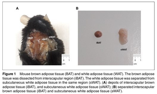
Adipocytes play a critical role in energy metabolism. White adipocytes store energy in a single large lipid droplet, while brown adipocytes contain multiple small lipid droplets and many mitochondria, contributing to non-shivering thermogenesis, which is crucial for maintaining body temperature. Due to its thermogenic properties in response to cold and beta-adrenergic agonists, activating brown adipose tissue, which was unexpectedly discovered in adult humans through radiological approaches, has become an attractive strategy for maintaining energy homeostasis and treating metabolic disorders.
Beyond their roles in metabolic processes, adipocytes secrete many cytokines, also known as adipokines. Both white and brown adipocytes secrete common cytokines, such as adiponectin and IL-6, but they also secrete distinct sets of cytokines.
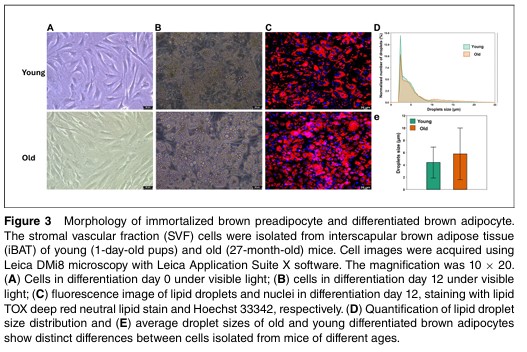
Brown and white adipocytes also play significant roles in aging. As individuals age, the function and thermogenic capacity of brown adipocytes decline, slowing metabolic rates and increasing the risk of insulin resistance, obesity, type 2 diabetes and other metabolic disorders. Meanwhile, white adipocytes persistently secrete pro-inflammatory cytokines, such as TNF-α, IL-6, and MCP-1, leading to a state of chronic low-grade inflammation that contributes to various age-related diseases, including cardiovascular diseases and certain cancers.
To better understand and investigate the roles of brown and white adipocytes in energy homeostasis, thermogenesis, metabolic disorders, and aging processes in vitro, we established immortalized mouse brown and white preadipocyte cell lines from young (1-day old pups and 20-week-old) and old mice(12-month-old and 27-month-old).One group of old mice (12 months) was also maintained on a normal diet and a high-fat, high-fructose diet for 10 months.
Biosafety Level
BSL2
Culture Medium
449 ml Dulbecco’s modified Eagle medium/F12 (DMEM/F12) (Cytiva, cat. no. SH30023.01) 50 ml FBS (Gemini Bio, cat. no. 100-106) 1 ml of primocin (Fisher, cat. no. NC9141851) Filter medium through 0.2-μm filters (Fisher, cat. no. FB12566510). Store up to 6 months at 4°C DMEM/F-12 medium containing 10% FBS and 100 μg/ml primocin.
Depositor
Dr. Sui-Seng Tee Lab at UMBC
Freeze Medium
10 ml dimethyl sulfoxide (DMSO) (Sigma, cat. no. D2650) 90 ml fetal bovine serum (FBS) (Gemini Bio, cat. no. 100-106) Filter mixture with a 0.2-μm filter (Fisher, cat. no. FB12566510) Store up to 1 year at −80°C
Growth Properties
Adherent
Strain
Male C57/BL6
Tissue
Adipose Tissue
Species
Mouse
Age At Sampling
12-month-old
Applications Range
Study of energy homeostasis, thermogenesis, metabolic disorders, and aging processes
Shipping
Shipped on dry ice
Product Format
Frozen
Thawing Protocol
1. Take cryotubes containing preadipocytes out of a liquid nitrogen tank. 2. Keep cryotubes in a 37°C water bath for 2 to 3 min to thaw the cells. 3. Centrifuge cryotubes 3 min at 400 × g,4°C. 4. Discardsupernatant (freezing medium) and do not disturb the cell pellet in the bottom of the tube; the cell pellet could be visible or invisible. 5. Add 1mlcell growth medium into each cryotube, and gently vortex for 10 s. 6. Transfer the 1 ml cell suspension to a new 50-ml tube. 7. Add additional cell growth medium and gently vortex for 10 s. 8. Plate the cells in tissue culture plates, dishes, or flasks. To yield 40% to 50% confluence, seed 1 × 10⁵ cells into one well of 6-well plate; 1 × 10⁶ cells into one 10-cm dish; 1.5 × 10⁶ cells into one T-75 flask. 9. Culture the cells in growth medium at 37°C in a humidified incubator with 5% CO2.
Freezing Protocol
1. Wash preadipocytes with 1× PBS for one time and add a couple of drops of trypsin to cover the cell surface. 2. Incubate the cells at 37°C in the incubator for 2 to 3 min. 3. Add growth medium to the well (1 ml/well, 6-well plate). 4. Detach and mix cells by pipetting up and down. 5. Collect cells in a new 50-ml tube. 6. Centrifuge the cells 3 min at 400 × g,4°C. 7. Carefully remove the supernatant and avoid disturbing the cell pellet. 8. Add cell freezing medium to the tube and vortex for 30 s to resuspend cells. Usually, 3 ml freezing medium is used for the cells collected from one plate (6-well plate). 9. Aliquot 1 ml cells in a 1 ml cryotube. 10. Place cell cryotubes in a freezing container containing 100% isopropyl alcohol and keep them in a–80°C freezer overnight. 11. Store the cells in liquid nitrogen for long-term preservation.
Subculture Protocol
When the preadipocytes reach 80% to 90% confluence, subculture cells with a ratio of 1:3 to 1:4, which will yield 30% to 40% confluence.
Materials Preadipocytes (see Support Protocol 2 or Basic Protocols 3, 4, and 5) 1×PBS, calcium- and magnesium-free (Fisher, cat. no. 10-010-049) 0.25% trypsin-EDTA (1×) (Gibco, cat. no. 25200-056) Primary preadipocyte growth medium (see recipe) Cell culture incubator, 37°C, 5% CO2 Inverted microscope 50-ml Falcon tubes (Corning, cat. no. 352070) Tabletop centrifuge Vortexer Tissue culture plates, dishes, or flasks
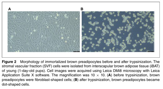
Procedures: 1. Aspirate culture medium, wash cells once with 1× PBS. 2. Add 0.25% (w/v) trypsin-EDTA to cover the cell surface. 3. Incubate the cells in an incubator at 37°C for 2 to 3 min. 4. Observe cells under an inverted microscopy with 4× or 10× magnification. When fibroblast-shaped preadipocytes shrink and become dot-shaped cells (Fig. 2), add growth medium to inactivate trypsin (1 ml/well of a 6-well plate). 5. Detach cells by pipetting up and down, and then collect cells to a new 50-ml tube. 6. Centrifuge the cell suspension 3 min at 400 × g, room temperature. 7. Carefully discard the supernatant (trypsin and medium), do not disturb the cell pellet in the bottom of the tube. The cell pellet may be visible or invisible. 8. Add growth medium to the tube, and vortex for 10 s. Usually one 6-well plate of preadipocytes can split into three to four 6-well plates; add growth medium accordingly. 9. Plate the cells in new tissue culture plates, dishes, or flasks. 10. Culture the cells in the incubator at 37°C, with 5% CO2, to allow the cells to grow. 11. Replace with fresh growth medium every other day. When the cells reach 80% to 90% confluence, either subculture cells again or freeze cells for cryopreservation.
No Description Available.
No Description Available.
Reviews of Immortalized Old Mouse Brown Preadipocyte Cell Line
 Loading ....