 Loading ....
Western Blot Imaging System
Lens-Free, Contact-Based Western Blot Imaging — Clarity in 1 Second
Capture faint and strong bands in a single exposure. Streamlined workflow, ultra-high sensitivity, and expanded dynamic range—purpose-built for reproducible protein quantification.
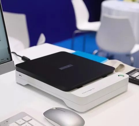
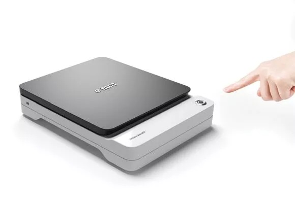
E-BLOT Touch Imager — compact, contact-based instrument.
Why contact-based imaging changes everything
Touch Imager places the membrane directly on an oversized CMOS sensor—eliminating lenses and signal loss. The result: faster imaging, higher sensitivity, and accurate quantification without saturation.
Ultra-high sensitivity
Capture low-abundance targets and reduce repeats.
Expanded dynamic range
Faint and saturated bands in a single exposure.
1-second capture
Press once. Get results. No manual tuning.
90% smaller footprint
All-in-one form factor frees bench space.
Imaging Architectures
How signal travels in different systems.
Compare imaging approaches
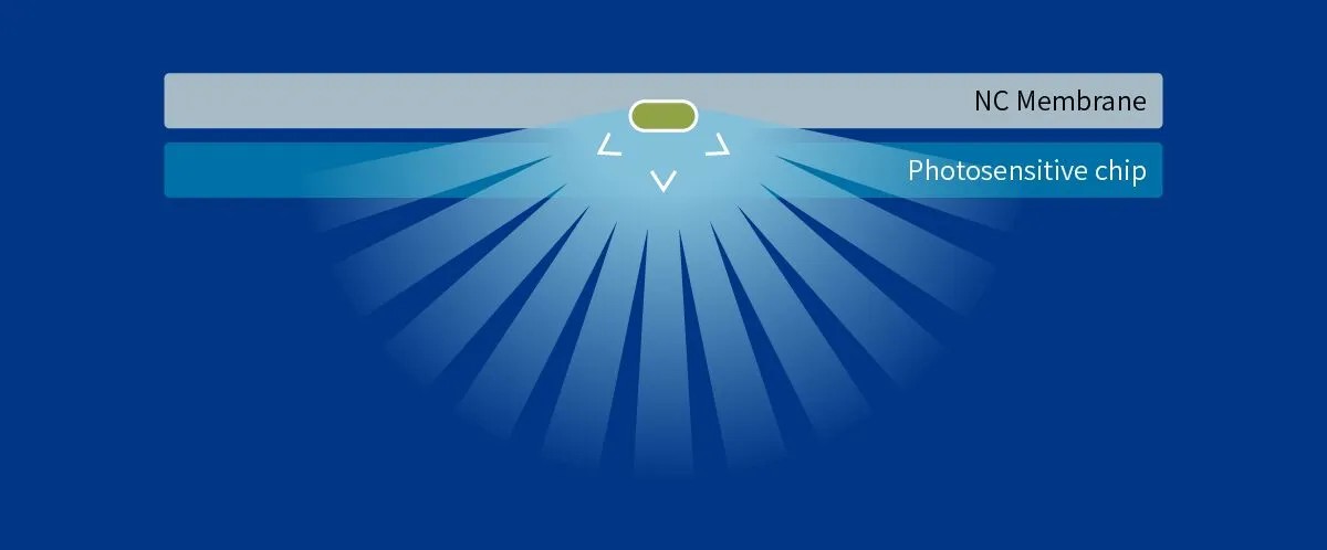
Touch Imager (Contact-based)
- High sensitivity and SNR without optical loss
- Single-shot capture of weak & strong bands
- 1-second imaging, no exposure tuning
- Compact, no darkroom or bulky optics
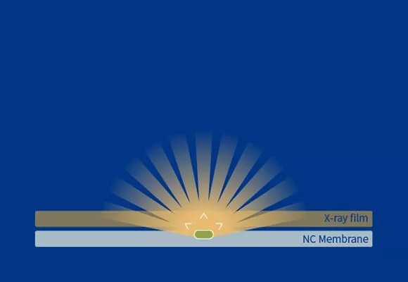
X-ray film
- High maintenance & consumables
- Narrow dynamic range; repeat exposures common
- Uses toxic processing chemicals
- Darkroom setup and long workflows
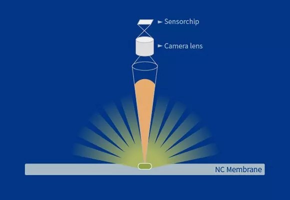
Cooled CCD
- Significant optical signal loss
- Limited dynamic range; saturation risks
- Longer exposures for similar clarity
- Bulky optics and larger footprint
The Imaging Technology That Performs Better In All Scenarios
Touch Imager vs. X-ray Film: Superior clarity with minimal exposure time
1.jpg)
α-Tubulin Testing (Left Panel)
Touch Imager shows uniform, crisp bands with high clarity and contrast in a 1-second exposure, minimizing re-runs and preserving quantitation.
The X-ray film control (typical 30-second exposure) needs longer exposure to reveal faint bands and risks saturation in high-intensity lanes.
Protein Sample Testing (Right Panel)
Multiple proteins are cleanly resolved in a 1-second capture on Touch Imager with excellent uniformity and low background.
The X-ray film comparison (typical 60-second exposure) shows higher background and band broadening.
1.jpg)
Touch Imager vs. Cooled CCD: Faster imaging with enhanced clarity
.jpg)
α-Tubulin Testing (Left Panel)
Touch Imager provides high-contrast, uniform bands in a 1-second capture, ideal for quantitation without manual tuning.
The cooled CCD comparison (typical 30-second exposure) shows reduced contrast and uneven band intensities, especially at the edges.
Protein Sample Testing (Right Panel)
Multiple proteins remain clearly defined with strong SNR in a 1-second capture on Touch Imager.
The CCD comparison (typical 60-second exposure) shows broader bands and higher background with longer exposures.
.jpg)
Technical highlights
| Imaging method | Contact-based, lens-free CMOS |
|---|---|
| Capture time | ~1 second (typical) |
| Sensitivity | Optimized for low-abundance targets |
| Dynamic range | Expanded; strong & weak bands in one shot |
| Footprint | Compact, bench-friendly |
| Use cases | Western blot, quantitative band analysis |
Representative results and device details.
Frequently asked questions
Request a demo
Tell us about your workflows and we’ll tailor a live session to your use case.
Talk to an expert
Prefer a quote path? Use our contact flow and we’ll respond within one business day.