 Loading ....
A53T Alpha Synuclein in Parkinson’s Disease

A53T Alpha Synuclein in Parkinson’s Disease

Copyright © StressMarq Biosciences Inc 2018-2019 All rights reserved
Alpha Synuclein in Parkinson’s Disease
Parkinson’s Disease (PD) is a neurodegenerative disorder characterized by the aggregation of alpha synuclein into Lewy bodies in the brain. Alpha synuclein is a 14-kDa protein encoded by the SNCA gene. There is conflicting evidence regarding the native state of alpha synuclein: it may be an unfolded monomer,1 folded tetramer, or a dynamic equilibrium between these and other oligomers.2 In Parkinson’s Disease, these smaller forms aggregate into protofibrils, fibrils, and ultimate Lewy bodies, which are thought to cause neuronal dysfunction and death. There is also the possibility that the oligomers and protofibrils are the neurotoxic species3-6 while Lewy bodies are neuroprotective.7
What is the A53T mutation?
A53T is a missense point mutation, meaning one amino acid is changed: the 53rd amino acid is changed from alanine to threonine. This mutation is caused by guanine being changed to adenine at position 209 of the SNCA gene (G209A).8
What is the role of the A53T mutation in Parkinson’s Disease?
The A53T mutation has been linked to autosomal dominant, early-onset PD, first documented in families of Italian and Greek descent.8 It has also been documented in a Korean family.9 A53T mutation results in an earlier age of onset and a shorter disease duration.8 Even though most cases of PD are sporadic, not inherited, and do not involve the A53T mutation, understanding the role of A53T could help researchers better understand mechanisms of alpha synuclein aggregation and develop better models and therapies.
A53T Aggregation
A53T alpha synuclein fibrillizes in solution faster than wild-type (WT) alpha synuclein.10 The mutation, however, has a greater effect on the rate of alpha synuclein protofibril formation than on the rate of fibril elongation.11 The fact that A53T and A30P mutations are both linked to early-onset PD and promote protofibril formation suggests that protofibrils contribute to the neurodegeneration seen in PD.12
Why does A53T alpha synuclein fibrillize faster than WT alpha synuclein?
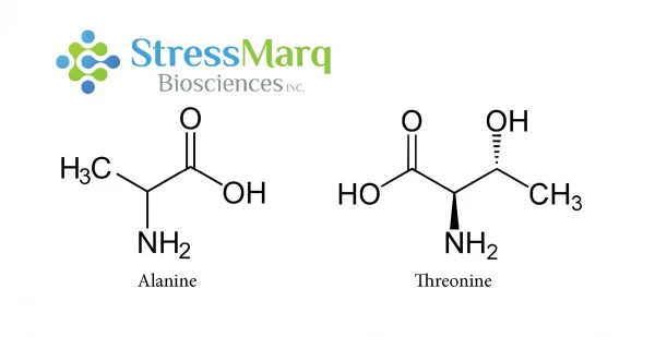
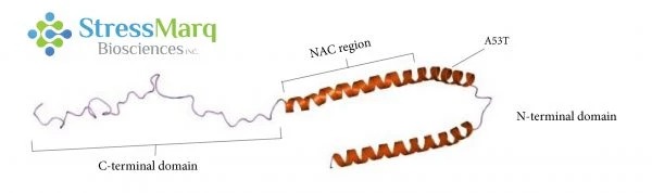
The A53T mutation only involves a single changed amino acid, and alanine and threonine are structurally similar, so why is there such a significant effect? A53T and other disease-causing mutations occur in the N-terminal region of the alpha synuclein protein. Theoretical models indicate that the A53T mutation causes long-range interactions between the NAC region and the N- or C-terminal regions of alpha synuclein to disappear, leading to an increase in beta-sheet formation.13 NMR measurements indicate that the A53T mutation could extend and stabilize beta-sheet structures involved in oligomerization and fibrillization.14
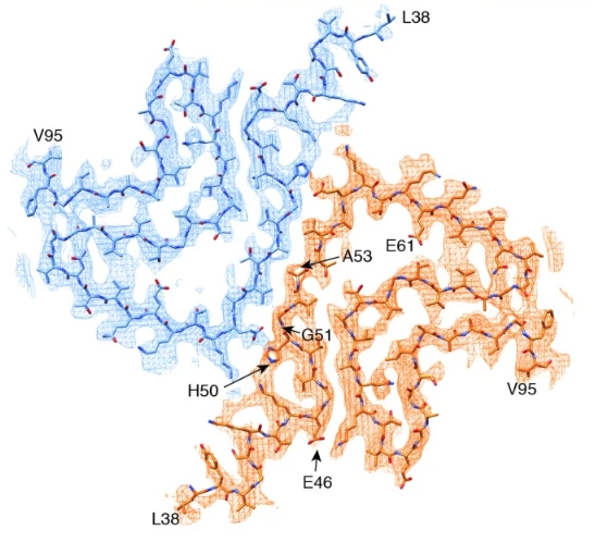
Cryo-EM analysis revealed that alpha synuclein fibrils consist of two protofilaments twisted together into left-handed helices.16 The A53T mutation, along with four of the five other early-onset PD mutations, is located at the interphase between the two protofilaments.16 Introducing a hydrophilic residue such as threonine disrupts this “steric-zipper interphase” and changes the structure of the fibrils.16
Effects of the A53T Mutation
Regardless of the mechanism of A53T alpha synuclein aggregation, it has devastating downstream effects. A53T alpha synuclein expression in mice leads to motor deficits, paralysis, and death.17 A53T alpha synuclein has a greater tendency than WT alpha synuclein to form annular and tubular protofibrils, and A53T alpha synuclein protofibrils bind tightly to phospholipid vesicles, leading to their permeabilization.12 These lipid vesicles can induce fibril production by enhancing nucleation of monomers.18 The A53T mutation is thought to promote fibrillization by either changing the structure of vesicle pores or by promoting the formation permeabilizing protofibrils over other species.19
Dopamine
Dopamine is a neurotransmitter that regulates speech and movement. Parkinson’s Disease motor deficits are caused by a loss in dopamine-secreting (dopaminergenic) neurons. Mice overexpressing A53T alpha synuclein experience impaired dopamine signaling prior to neurodegeneration,20 and rats expressing A53T alpha synuclein showed greater losses of dopaminergic neurons than their counterparts expressing WT alpha synuclein.21 A53T alpha synuclein expression is also thought to make cells more vulnerable to dopamine toxicity.22
Synapses
Synapses are components of neurons responsible for the transfer of information between neurons via synaptic vesicles.23 Both A53T and WT alpha synuclein suppress presynaptic transmission by depleting the synaptic vesicle recycling pool.24 A53T alpha synuclein expression also produces postsynaptic deficits.25 These postsynaptic deficits are caused by the hyperphosphorylation of tau and its mislocalization to dendritic spines.24
Microglia
Microglia are responsible for synaptic pruning and respond to injuries by producing inflammatory mediators.26 Activated microglia change morphologies, produce reactive oxygen species (ROS), and release inflammatory cytokines.27 In Parkinson’s Disease, they may become overactivated and contribute to the depletion of dopaminergic neurons.27 A53T alpha synuclein activates microglia more strongly than WT or other mutants, leading to microgliosis and a subsequent inflammatory state.27 A53T alpha synuclein expression also makes cells more vulnerable to oxidative stress.28
Astrocytes
When A53T alpha synuclein is selectively expressed in mouse astrocytes, it leads to rapid paralysis.29 This is thought to be due decreased glutamate transporter expression disruption leading to downstream activation of microglia.29
Mitochondrial Dysfunction and Endoplasmic Reticulum Stress
A53T alpha synuclein-induced inflammation and oxidative stress affect both the mitochondria and the endoplasmic reticulum (ER). A53T expression causes mitochondrial dysfunction involving impaired mitochondrial membrane potential and respiratory function.30 This in turn leads to mitochondrial autophagy.31 Overactivation of autophagy may lead to the mitochondrial loss and resultant neurodegeneration.31 However, it may also have some protective effects by clearing aggregated proteins.31 A53T neurons showed impaired mitochondrial motility that could be reversed by rapamycin, an autophagy inducer.30
In differentiated PC12 cells, increasing A53T alpha synuclein expression led to up to 40% cell death due to increased intracellular ROS level and decreased proteasome activity.32 In this case, ER stress and mitochondrial function involved the release of mitochondrial cytochrome C and elevated caspase-3, -9, and -12 activities, and led to cell death.32
Therapeutic Approaches
Preventing the neurodegeneration associated with A53T mutation could lead to potential PD therapies. A COX-1 inhibitor extends the lifespan of A53T alpha synuclein-expressing mice,29 and Cyclosporin A, Z-Vad, and Caspase-3 and -9 inhibitors reduced cell death from ER and mitochondrial stress.32 Although the A53T mutation is not present in the majority of PD cases, WT alpha synuclein may have similar but slower mechanisms of fibrillization and subsequent neurodegeneration. A53T mutated alpha synuclein was more effective than WT alpha synuclein in establishing a PD model in rats, and resulted in a model representative of early-onset PD.21 An enhanced understanding of A53T alpha synuclein fibrillization and inhibition mechanisms could help those with the A53T mutation in addition to other PD patients.