 Loading ....
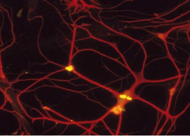
Studies have found that Miller glial cells can regenerate nerve cells in the retina

Studies have found that Miller glial cells can regenerate nerve cells in the retina

Some cells in the human eye can repair the damage caused by visually impaired diseases. But so far, scientists have not succeeded in making them work.
Now, a research team claims to have motivated these cells -- called Miller glial cells -- to regenerate a photoreceptor cell in the eyes of mice.
The study found that these new cells can detect the incoming light and form a network with other cells in the eye to transmit signals to the brain, which is a potential step to reverse some genetic eye diseases and injuries, which is also used to treat retinal pigments. Blind diseases such as degeneration bring new hope.
The retina of the zebrafish has a self-repairing function. When the retina is damaged, the "Muller glial cells" can regenerate the nerve cells of the retina, but the mammalian cells do not have similar regeneration functions. Although the scientific community has been able to activate mammalian "Muller glial cells" by damaging the retina, this method is more harmful to the retina and is not conducive to restoring vision.
The researchers used gene transfer methods in mouse experiments to cause Miller glial cells to divide and develop into sensitized rod cells. The newly developed rod cells are structurally indistinguishable from natural rod cells and form synaptic structures that allow them to communicate with other nerve cells in the retina. Experiments have shown that this method can restore the blind mice.
This is the first time scientists have reprogrammed Miller's glial cells into functioning rods in the mammalian retina. Rod cells allow people to see things in low light conditions and may help protect cones, which are responsible for distinguishing colors and improving visual acuity.
No one can create a cell like a photoreceptor like them. Even in mice that regenerated the most new rod cells, the density was only 0.2% of the retina of healthy mice. As a result, the treated mice are able to perceive the light, but they cannot recognize the shape or the object.
Research has cracked the first part of the problem, and the problem now is to enlarge it. If the study allows Miller's glial cells to produce more photoreceptor cells, this approach may one day restore some of the vision of those who lose their rods because of retinal detachment or hereditary retinitis.
The research team still needs to prove that rod cell development and function are normal in the diseased eye - the retinal cells here may not connect and interact properly.
In blind mice, existing rod-shaped cells are repaired during surgery, either because they receive the correct genes carried by the virus, or because Miller's glial cells share the correct genes with them. . In both cases, the brain's visual signal is not derived from newly generated rod-shaped cells, but from the recovery of existing photoreceptor cells. This study ignores the chemical labeling technique, which can prove that any functional rod cell is derived from Miller glial cells.
This research team has done such a labeling experiment, and the origin of new rod cells has been thoroughly proved by several other methods. A control experiment was also cited, in which the team transferred the corrective gene to Miller's glial cells without reprogramming. In this case, there is no visual signal in the brain, which means that the existing rod cells are not recovered.