 Loading ....
The study found that a three-pronged treatment can trigger axonal regeneration after spinal cord injury

The study found that a three-pronged treatment can trigger axonal regeneration after spinal cord injury

Copyright © iCell Bioscience Inc, Shanghai 2018-2019
When people suffer from spinal cord injury, this can damage the axons and prevent the brain from sending signals to neurons beneath the injury site, leading to loss of paralysis and other neurological functions such as bladder control and hand strength. Axons are tiny nerve fibers that connect our neurons and enable them to communicate.
In a new study, researchers from the University of California, Los Angeles, Harvard, and the Swiss Federal Institute of Technology identified a three-pronged treatment that triggers axonal to undergo complete spinal cord in rodents. It can be regenerated after injury.
Decades of research have shown that our nerve fibers require three processes to grow: first, to initiate axonal growth through genetic programming; second, molecular pathways on which fiber capture and growth depend; and third, to attract axons along A protein "breadcrumb" trajectory that grows in a particular direction.
These three processes are active when a person develops in the womb. After birth, they are turned off, but the genes that control the growth process are still asleep in our bodies. Re-awake these genes and then restart the process in a three-pronged approach.
First, the researchers reactivated nerve cells in the spinal cord of mice by injecting osteopontin, insulin-like growth factor 1 and ciliated neurotrophic factors in a harmless virus. Two weeks later, the researchers anesthetized the mice and lost their connection between the axons in the lower part of the spinal cord. Only their hind legs are affected, but they are still able to move and eat.
Two days after the injury, the researchers used fibroblast growth factor 2 = and epidermal growth factor for a second treatment of the lesion to create a new pathway that is more suitable for axonal growth. Finally = release of a third group of molecules known as chemoattractant---glial cell-derived neurotrophic factor =---. The axons look for these chemical "bread crumbs", where the chemical "bread crumbs" provide the target location. For the purposes of this study, the target location refers to the spinal tissue present on the other side of the injury site.
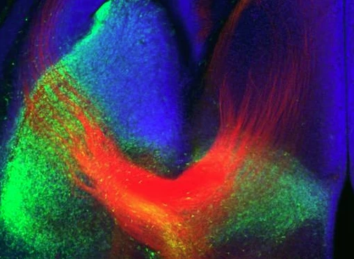
When the researchers examined the spinal cord tissue of mice treated with this three-pronged approach, they found that not only axonal growth through scar tissue, but also many nerve fibers have penetrated into the spinal cord tissue on the other side of the injury site. The neurons there have established new connections. None of the mice receiving this combination showed axonal regeneration in the injured area.
In order to test the reproducibility of these findings, repeated experiments were performed on rats, and robust results were also obtained. New findings were obtained when testing whether newly regenerated axons could perform electrical activity in these living animals.
When the spinal cord of these animals is stimulated with low current above the injury site, these regenerated axons transmit 20% of normal electrical activity below the injury site. In contrast, animals that did not receive treatment did not show this.
Although these findings suggest that these newly formed connections are capable of transmitting signals across the site of injury, the mobility of these rodents has not improved.
These regenerated axons act like axons formed during development, but they do not immediately support coordination. Just as newborns must learn to walk, axons that regenerate after injury will require training and practice before they can resume function.
The researchers then sought to explore how to retrain these newly formed neural connections in order to restore the movement of these animals.