 Loading ....
Research and development of a new method for analyzing cells and their components - immunofluorescence imaging

Research and development of a new method for analyzing cells and their components - immunofluorescence imaging

Copyright © iCell Bioscience Inc, Shanghai 2018-2019
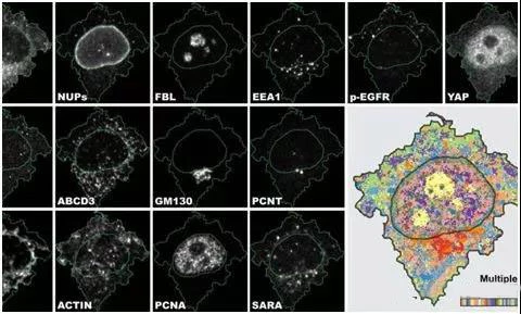
In a new study, researchers from the University of Zurich (UZH) in Switzerland developed a new method for analyzing cells and their components, namely iterative indirect immunofluorescence imaging (4i). This innovative approach greatly improves the standard immunofluorescence imaging techniques used in biomedical applications and provides clinicians with a wealth of data from each sample.
4i makes it possible to simultaneously observe the spatial distribution of at least 40 proteins and their modifications in the same cell in hundreds of thousands of cells at different levels from tissue to organelle.
4i is the first imaging technology to provide multiple observations of biological samples at different levels from tissue to organelles. It is possible for the first time to correlate multiple information obtained at the tissue, cell and subcellular levels in the same experiment.
Immunofluorescence (IF) uses antibodies to visualize and localize proteins in biological samples. Although the standard IF method typically labels three proteins, 4i visualizes more than ten times more protein by iterative hybridization and removal of antibodies in the sample using off-the-shelf antibodies and conventional fluorescence microscopy.
Once you have access to this large amount of data, you must analyze them - this is the next challenge for these researchers. Subcellular resolution images of thousands of cells were obtained over 10 channels under 10 processing conditions. The human eye and brain cannot handle the complex biological data collected by 4i.
To take full advantage of 4i data, Gut developed a new computer program for visual observation and analysis, a multiplexed protein map. It extracts multiple fluorescent signals from millions of pixels and produces an abstract but representative multiplexed protein profile in the cell.
Therefore, these researchers were able to systematically investigate cell landscapes: they successfully visualized the intracellular spatial distribution of most mammalian organelles in the cell cycle and in different environments.
The application of 4i and multiplex protein maps is multifaceted, from basic research to precision medicine. It is hoped that 4i and multiple protein maps will help scientists better understand the process that has been at the center of biological research for decades.
At the same time, these researchers plan to use these technologies to advance the development of precision medicine, especially in cancer diagnosis and treatment options. This 4i assay can also be used to determine the effect of pharmacologically active substances on cell distribution and physiological properties. It is currently being used by clinicians and pharmaceutical companies in translational research to improve the treatment outcomes of cancer patients.
The research team wanted to describe the tumor cell characteristics of patients who had been treated with different cancer drugs. These researchers hope that these laboratory results will inform clinical decisions that support individualized treatment of patients. In addition, 4i and multiplex protein profiling on tumor tissue sections is planned to identify relevant biomarkers to improve diagnosis and prognosis for cancer patients.