
Description
Protein A/G MagBeads use "nano-surface biotechnology" (S-TEC) to target Protein A/G onto the surface of superparamagnetic polymer microspheres with high density. It has higher antibody binding ability and very low non-specific adsorption rate of protein. One-step purification can isolate the antibody with purity >90% from the serum sample, which is simple and effective to use. Natural Protein A is a cell wall surface protein found in Staphylococcus aureus. Natural Protein G is a cell surface protein isolated from the genus G or C Streptococcus. Both have similar functions and bind most mammalian IgG by interacting primarily with the Fc region of immunoglobulin (Ig), but differ in their binding specificity. Protein A/G MagBeads covalently conjugate both protein A and protein G, providing A wider binding range and higher utility than either protein A or protein G alone. At the same time, this product uses genetically modified proteins A and G, which not only maintain their Ig affinity properties, but also remove the non-major binding domain of the natural protein itself to reduce non-specific binding. This product has a wide range of application, can be used in cell lysis fluid, cell secretory fluid supernatant, serum, animal ascites and other immune antigen samples.
Yeasen offers comprehensive solutions for protein purification experiments. Explore our related products: Antibody Capture Tool: Easily Handle IP and Co-IP with Protein A/G Agarose and Magnetic Beads
Features
- Low background—little to no nonspecific binding, and no preclearing
- Highly reproducible—uniform beads ensure the most consistent results
- Highly sensitive—Magbeads technology is the most cited method for sensitive applications, such as ChIP and IP, of low abundance proteins
- Fast and easy— no centrifugation or preclearing steps
- Versatile—products for IP, Co-IP, pull-down, and ChIP assays.
Applications
- Immunoprecipitation (IP)
- Purification of antibody
Specifications
| Ligand | Protein A/G |
| Binding Capacity | ≥50 μg hIgG /mg |
| Particle size | 1 μm |
| Concentration | 10 mg/mL |
| Storage Buffer | PBS, 0.01% Tween-20, 0.02% NaN3 |
Smaller beads, Higher capacity
|
Supplier T* Protein G/Protein A |
Yeasen Protein A/G MagBeads, 36417ES |
|
|
Diameter |
2.8 μm |
1 μm |
|
Concentration |
30 mg/mL |
10 mg/mL |
|
Capacity |
About 8 µg human IgG/mg beads |
About 50µg human IgG /mg beads |
Components
| Components No. | Name | 36417ES03 | 36417ES08 |
| 36417 | Protein A/G MagBeads (IP Grade) | 1 mL | 5 mL |
Storage
The products should be stably stored at 2~8℃ for 2 year.
Figures


 Cited from: Oncogene. 2022;41(10):1482-91
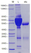
The SDS-PAGE gel electrophoresis results of Rabbit serum purification by Protein A/G MagBeads (IP Grade).L: sample, L: eluate.
Documents:
36417_Manual_HB230112_EN.pdf
Citations & References:
[1] Zhou L, Zhuang H-J, Chen Q, Jiang L-P, Han X-M, Ge Y-X, et al. Precise targeting of osteopontin in non-small cell lung cancer spinal metastasis to promote chemosensitivity via a smart hollow nano-platform. Chemical Engineering Journal. 2022;436:132131.(IF:15.1)
[2] Jiang Y, Guo H, Tong T, et al. lncRNA lnc-POP1-1 upregulated by VN1R5 promotes cisplatin resistance in head and neck squamous cell carcinoma through interaction with MCM5. Mol Ther. 2022;30(1):448-467. (IF:11.454)
[3] Wang Y, Gao W-Y, Wang L-L, Wang R-L, Yang Z-X, Luo F-Q, et al. FBXW24 controls female meiotic prophase progression by regulating SYCP3 ubiquitination. Clinical and Translational Medicine. 2022;12(7):e891.(IF:10.6)
[4] Wang Y, Gao WY, Wang LL, et al. FBXW24 controls female meiotic prophase progression by regulating SYCP3 ubiquitination. Clin Transl Med. 2022;12(7):e891. doi:10.1002/ctm2.891(IF:8.554)
[5] Cen Y, Zou X, Zhong Q, et al. The TIAR-mediated Nrf2 response to oxidative stress is mediated through the Nrf2 noncoding 3'untranslated region in Spodoptera litura. Free Radic Biol Med. 2022;184:17-29. doi:10.1016/j.freeradbiomed.2022.03.016(IF:7.376)
[6] Hua Z, Wei R, Guo M, Lin Z, Yu X, Li X, et al. YTHDF2 promotes multiple myeloma cell proliferation via STAT5A/MAP2K2/p-ERK axis. Oncogene. 2022;41(10):1482-91.(IF:8.0)
[7] Zhou W, He P, Liu H, Wei H, Yu J. A luciferase based automated assay for rapid and sensitive detection of SARS-CoV-2 antibodies. Analytica Chimica Acta. 2023;1238:340633.(IF:6.2)
[8] Zhenzhen L, Wenting L, Jianmin Z, et al. miR-146a-5p/TXNIP axis attenuates intestinal ischemia-reperfusion injury by inhibiting autophagy via the PRKAA/mTOR signaling pathway. Biochem Pharmacol. 2022;197:114839. doi:10.1016/j.bcp.2021.114839(IF:5.858)
[9] Wang L, Yi J, Yin XY, et al. Vacuolating Cytotoxin A Triggers Mitophagy in Helicobacter pylori-Infected Human Gastric Epithelium Cells. Front Oncol. 2022;12:881829. Published 2022 Jul 14. doi:10.3389/fonc.2022.881829(IF:5.738)
[10] Meng J, Zhang C, Wang D, Zhu L, Wang L. Mitochondrial GCN5L1 regulates cytosolic redox state and hepatic gluconeogenesis via glycerol phosphate shuttle GPD2 [published online ahead of print, 2022 Jun 28]. Biochem Biophys Res Commun. 2022;621:1-7. doi:10.1016/j.bbrc.2022.06.092(IF:3.575)
[11] Chen P, Wang L, Long YB, et al. E2F4 regulates the cell cycle and DNA replication in the silkworm, Bombyx mori. Insect Sci. 2022;29(4):1006-1016. doi:10.1111/1744-7917.12991(IF:3.262)
S-TEC (Nano-Surface Biotechnology) is a surface engineering method that targets Protein A/G onto the magnetic bead surface with high density in a controlled orientation. This maximizes the number of accessible Fc-binding sites per bead compared to conventional random coupling methods. The result: higher IgG binding capacity (≥50 µg hIgG/mg beads) and lower non-specific protein adsorption than competing 2.8 µm beads at 30 mg/mL concentration.
Protein A/G is a fusion of Protein A and Protein G that combines the binding advantages of both: it binds human IgG1–4, mouse IgG1–3, rat, rabbit, goat, horse, and cow IgG. Mouse IgG1 and human IgG3 bind well to Protein G (and therefore Protein A/G) but weakly to Protein A alone. Check your antibody's species and subclass before use.
Add the serum sample (diluted 1:1–1:10 with PBS) directly to pre-equilibrated Protein A/G MagBeads in binding buffer (PBS, pH 7.0–8.0). Incubate 30–60 min at room temperature or 4°C with rotation. Wash 3× with PBS. Elute with 100 mM glycine–HCl, pH 2.5–3.0, and immediately neutralize with 1 M Tris-HCl, pH 9.0. Measure purity by SDS-PAGE or SEC.
Yeasen Protein A/G MagBeads are 1 µm in diameter (vs. competitor 2.8 µm) and at 10 mg/mL (vs. 30 mg/mL). Smaller size provides greater surface area per volume and better in-solution suspension kinetics for faster antibody capture from dilute samples. Binding capacity (≥50 µg hIgG/mg beads) substantially exceeds typical 2.8 µm formats (~8 µg/mg).
Store at 2–8°C for up to 2 years. Do not freeze. Storage buffer: PBS, 0.01% Tween-20, 0.02% NaN₃. Ensure complete bead resuspension by vortexing before use. Magnetic beads settle during storage — always mix immediately before pipetting.
The following customization and add-on services may be available for this product type through the supplier. For inquiries and pricing, contact support@biohippo.com.
Customization Options
- Bulk Quantities: Larger volumes and custom pack sizes beyond the standard catalog may be available for high-throughput core facilities and industrial customers. Contact us to inquire about bulk pricing.
- Custom Antibody Coupling (Magnetic Beads): Custom magnetic beads conjugated to your specific antibody or protein ligand may be available through Yeasen's OEM and custom services program. These services leverage the same bead platform used for catalog products.
- OEM Manufacturing: Custom bead formulations for diagnostic device development, including magnetic beads for nucleic acid isolation, IP-grade beads, and affinity capture beads, may be available at manufacturing scale.
To inquire about custom options or bulk pricing, contact support@biohippo.com.

