

| Field | Specification |
|---|---|
| Applications | |
| Production system | |
| Function | |
| Transgene | |
| Promoter | |
| Reporter/Tag | |
| Plasmid backbone | |
| Form | Liquid |
| Storage | |
| Storage buffer | |
| Shelf life | |
| Catalog no. (Mfr.) | |
| Main SKU |
Overview
Ad-Cre-IRES-GFP is a replication-defective recombinant Ad5 adenovirus expressing the Cre-IRES-GFP recombinase under the CMV promoter. It is used to deliver Cre activity to floxed alleles in cell lines and in vivo for conditional gene knockout, lineage tracing, and activation of Cre-dependent (DIO/FLEX) reporters.
Key elements and design rationale
- Backbone: Human adenovirus type 5 (Ad5) with E1 and E3 deleted (dE1/E3). Replication-incompetent in standard cells; replication-competent helper cells (HEK293) are required for amplification.
- Promoter (CMV): a strong, ubiquitous promoter active in most mammalian cell types.
- Transgene: Cre-IRES-GFP (eGFP tag).
- Titer & format: 1×10¹⁰ PFU/ml in storage buffer (DMEM, 2% BSA, 2.5% glycerol or equivalent), supplied as a 200 µL aliquot.
Biological background
Cre Recombinase is a Type I topoisomerase from bacteriophage P1 that catalyzes the site-specific recombination of DNA between loxP sites. loxP is a 34 bp DNA sequence at which confers directionality. Cre recombinase is used as a tool to genetically modify genes, such as to delete a segment of DNA flanked by LoxP sites in cells or experimental animals.
This adenovirus expresses both Cre recombinase and GFP. The GFP is in an IRES expression cassette.
Check out this instruction video on using our adenovirus Cre to knockout LoxP flanked gene in primary mouse embryonic fibroblasts (MEF).
Research relevance and current trends
- Decision-relevant for researchers studying Recombinases.
- Adenovirus-mediated delivery is well-established in primary cells, organoids, and small-animal models.
Common research applications
- Conditional gene knockout in floxed-allele cell lines and animals.
- Lineage tracing using Cre-dependent reporter alleles.
- Activation of Cre-dependent (DIO/FLEX) viral reporters or effectors.
Notes for experimental interpretation
- Recombination efficiency depends on Cre expression level, allele accessibility, and the time window assayed; allow 72–96 hours after infection for recombination to plateau.
- Adenoviral delivery is episomal and non-integrating; expression dilutes with cell division and typically lasts 1–2 weeks in dividing cells (longer in non-dividing cells such as hepatocytes, neurons, and cardiomyocytes).
- Pre-existing anti-Ad5 neutralizing antibodies are common in human and primate hosts and can reduce in vivo transduction; this is less relevant in inbred laboratory mouse strains.
- MOI optimization is essential — over-dosing can cause cytopathic effects; under-dosing yields incomplete transduction. A 3–5× MOI titration in your specific cell or animal model is recommended.
- Replication-defective Ad5 vectors are typically handled at BSL-2; consult your institutional biosafety officer for specific transgenes and routes of use.
The Adenovirus Genome
The adenovirus genome is a linear double-stranded DNA molecule of 26–46 kb that encodes 23–46 proteins (Figure 1). These genes are split into two functional categories:
- Early genes (E1–E4): encode proteins involved in viral transcription, viral DNA replication, and suppression of the host immune response.
- Late genes (L1–L5): encode viral capsid components and proteins required for capsid assembly.
These genes are flanked by inverted terminal repeats (ITRs) that initiate viral DNA replication and serve as binding sites for transcription factors.

Adenovirus Capsid and Serotypes
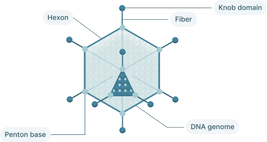
The adenovirus capsid is a non-enveloped icosahedral protein shell of approximately 70–90 nm in diameter, built from three major capsid proteins — hexon, penton base, and fiber — together with the minor proteins IIIa, VI, VIII, and IX (Figure 5). The 240 hexon trimers form the 20 triangular facets of the icosahedron, while 12 penton complexes occupy each vertex; each penton consists of a penton base anchored in the capsid and a trimeric fiber projecting outward. The fiber knob mediates initial attachment to host cell receptors, and the penton base then engages cellular integrins to drive internalization.
More than 50 human adenovirus serotypes have been characterized and are organized into seven species (A–G) based on hemagglutination, sequence homology, and receptor usage. Serotype determines tropism and primary receptor:
- Species C (e.g., Ad2, Ad5): bind the coxsackievirus and adenovirus receptor (CAR); broad tropism with strong transduction of liver and many epithelial cell types. Ad5 is the most widely used backbone in research and gene-delivery applications.
- Species B (e.g., Ad3, Ad11, Ad35): use CD46 as the primary receptor, giving access to cell types that express low levels of CAR.
- Species D: several members use sialic acid; some serotypes are associated with ocular tropism.
The replication-defective recombinant adenoviruses used as research vectors are typically derived from Ad5, with E1 (and often E3) deleted to render the virus non-replicative and to create space for transgene insertion.
What is this product?
Ad-Cre-IRES-GFP is a replication-defective recombinant Ad5 expressing Cre recombinase under the CMV promoter. It is used to deliver Cre activity to floxed alleles in cell lines and in vivo for conditional gene knockout, lineage tracing, or activation of Cre-dependent (DIO/FLEX) reporters.
How should I store and handle the virus?
Stocks are supplied at 1×10¹⁰ PFU/ml in storage buffer (typically 10 mM Tris pH 8.0, 2 mM MgCl2, 4% sucrose or similar). Store at −80 °C upon receipt, and aliquot before first use to minimize freeze-thaw cycles — recombinant adenovirus loses ~10–20% of infectious titer per freeze-thaw and should be limited to ≤3 cycles for quantitative work. Thaw on ice and dilute into pre-warmed culture medium immediately before infection.
Biosafety: Replication-defective Ad5 vectors are typically handled at BSL-2; consult your institutional biosafety officer for the specific transgene and route of use.
What MOI should I start with?
Optimal MOI varies by cell type, but useful starting ranges are:
- Most cell lines (HEK293, HeLa, U2OS, HepG2, etc.): MOI 10–100
- Primary cells (hepatocytes, cardiomyocytes, fibroblasts): MOI 50–500
- Resistant or low-CAR cells (some lymphocytes, hematopoietic): MOI 500–2000 (may give limited transduction; consider Ad5/35 fiber modification if available)
Worked example. To infect 1×10⁶ cells at MOI 100 from a 1×10¹⁰ PFU/ml stock: PFU needed = 100 × 106 = 1×10⁸ PFU; volume needed = 10 µl of stock. Always run a 3–5× MOI titration in your specific cell model to identify the dose that gives near-100% transduction without cytopathic effect.
When can I expect expression / activity?
Cre protein expression is detectable within 24 hours post-infection and reaches peak by 48–72 hours. Recombination at floxed alleles occurs continuously while Cre is expressed; allow 72–96 hours before assessing recombination efficiency to capture the full population.
What controls should I run?
For Cre delivery experiments, include:
- Ad-CMV-Null or Ad-CMV-GFP — capsid/dose control without Cre activity to control for any virus-induced effects.
- Cre-only (no floxed allele) control — cells of the same type lacking the floxed locus, infected with this vector, to confirm Cre itself does not cause the observed phenotype.
- Floxed allele without Cre — uninfected or mock-infected cells carrying the floxed locus, to confirm baseline expression of the floxed gene.
Can't find the adenovirus you need—or require a custom design and packaging service? We offer end-to-end adenoviral support for diverse research needs, including vector design and cloning, adenovirus construction for over-expression (from your plasmid, sequence, or RefSeq#), shRNA-silencing adenoviruses (from a working shRNA or via shRNA screening when you only have the target gene), and gRNA adenoviruses (from a gRNA cassette or sequence). Custom plasmid construction typically takes ~2 weeks, with viral packaging, purification, and QC adding another 2–4 weeks. Final stocks are CsCl-purified and PFU-titered, with deliverables of approximately 1×10¹² viral particles (1×10¹⁰–1×10¹¹ PFU). We also provide amplification and CsCl purification services at medium scale (~1×10¹⁰–5×10¹⁰ PFU/IFU in 2 mL, ~2 weeks; ideal for in vitro studies) and large scale (5×10¹²–1×10¹³ viral particles / 1–3×10¹¹ PFU, ~2–3 weeks; ideal for in vivo studies) — including amplification of customer-supplied viral stocks. Click Talk to a Scientist to submit a request, email us at support@biohippo.com, or explore our Research Services for additional support. Our team will be in contact with you shortly.

