

The Touch Imager open, with the 158 cm2 photosensitive chip exposed. e-BLOT states this field of view is 131 times the sensor area of a typical 1.2 cm2 cooled-CCD chip, so a full membrane is captured in one frame at 1:1 scale.

e-BLOT Touch Imager Western Blot Contact Imaging System
The e-BLOT Touch Imager is a lens-free western blot imaging system: instead of photographing the membrane through a camera lens, a 158 cm2 photosensor contacts the blot directly and records chemiluminescence at 1:1 scale. The manufacturer specifies that more than 95% of samples image within one second, with no detector cooling and no warm-up. A full well capacity of 1,250,000 electrons gives a wide intensity range, so strong and faint bands on the same blot can usually be captured in a single exposure. The instrument runs e-BLOT's Touch Viewer software for acquisition, image management, editing and densitometry, with multi-user accounts for shared-instrument labs. It is a 4.35 kg benchtop unit (27 x 20.6 x 5.4 cm) for western, Southern, Northern and dot blots and isotope imaging. Quoted individually; request a quote for pricing, lead time and demonstration availability.
| Catalog No. | Model | Availability | Price | Qty | Order |
|---|---|---|---|---|---|
| BHQ21100001 | Touch Imager | In Stock at Manufacturer | Quote | — |
Overview
What is e-BLOT Touch Imager Western Blot Contact Imaging System?
Overview
The Touch Imager is a benchtop chemiluminescence imaging system for Western blots that replaces the camera and lens of a conventional cooled-CCD imager with a large-format photosensor placed in direct contact with the membrane. The blot sits on the chip; the chip records the light where it is emitted. Nothing is focused, and nothing is collected across an air gap.
The practical consequence is speed and simplicity. e-BLOT specifies that more than 95% of samples are imaged within one second, and the instrument requires no detector cooling and no warm-up, so it is ready at power-on. A 158 cm² sensor covers a full membrane in a single frame, and a full well capacity of 1,250,000 electrons gives a wide intensity range in one exposure, so strong and faint bands on the same blot can usually be read without a bracketed exposure series.

How contact imaging differs from lens-based CCD imaging
A cooled-CCD imager forms an image the way a camera does: chemiluminescent photons leave the membrane in all directions, and only the small fraction that falls within the lens aperture reaches the sensor. Signal is therefore lost to distance and to the optics themselves, and the sensor is far smaller than the blot, so the image is a demagnified projection.

Contact imaging removes both losses. Because the photosensor is comparable in size to the membrane and touches it directly, photons are captured at 1:1 scale with no lens in the path. e-BLOT states that this yields sensitivity and quantitative range approximately two orders of magnitude beyond a cooled-CCD system. These are the manufacturer's figures; they have not been independently benchmarked by BioHippo, and we present them as manufacturer specifications rather than as our own measurements. The side-by-side exposures e-BLOT has published are reproduced in the Validation section below.

The trade-off is worth stating plainly: because imaging is contact-based and 1:1, the sensor area sets the maximum blot size, and the system images blots and membranes rather than serving as a general-purpose gel documentation camera for stained gels under transillumination.
Specifications
| Parameter | Specification |
|---|---|
| Detection principle | Direct contact imaging (lens-free) |
| Light sources | Chemiluminescence; epi-white illumination |
| Photosensor chip area | 158 cm² |
| Full well capacity | 1,250,000 electrons |
| Exposure time | >95% of samples imaged within 1 s |
| Capture modes | Automatic, Manual, Customized, Extreme HD |
| Start-up wait | None — no detector cooling required |
| Data transfer rate | 1000 Mbps |
| User management | Multi-user accounts |
| Dimensions (L × W × H) | 27 cm × 20.6 cm × 5.4 cm |
| Net weight | 4.35 kg |
| Power supply | 100–250 V |
| Operating temperature | 4–30 °C |
| Operating humidity | 10–85% relative humidity, non-condensing |
Source: e-BLOT published Touch Imager specifications. Confirm the current revision on your quotation before purchase.
Touch Viewer software
The instrument is operated through e-BLOT's Touch Viewer software, which covers the full workflow on one screen:
- Acquisition — automatic, manual, customized and Extreme HD capture, with saturation indication.
- Management — a gallery of saved images with review and sorting.
- Editing — rotate, flip, crop, and zoom, applied non-destructively to the saved original.
- Analysis — automatic or manual lane and band detection for densitometry.
- Accounts — multi-user login so images stay attributable in a shared-instrument lab.
Handling
The photosensitive chip is the imaging surface and is handled directly, so two points from the manufacturer's instructions matter in daily use. Use only the supplied plastic tweezers when placing and removing membranes — metal tweezers can damage the chip surface. Clean the chip with the supplied soft cloth. The instrument is calibrated before sample imaging, and the indicator lamp turns green when it is ready to capture.
For Research Use Only. Not for use in diagnostic procedures.
Details
Specifications
Applications
Applications & how to use
Western Blot (WB)
Detects a target protein in denatured, gel-separated lysates by antibody probing. Used to confirm protein identity, apparent molecular weight, and relative expression across samples.
Dot Blot
Protein or nucleic acid samples are spotted directly onto a membrane without electrophoretic separation, then detected with a labelled antibody or probe. Faster than a full blot when molecular weight information is not needed - used for antibody titration, screening large sample sets, and confirming target presence before committing to a Western blot.
Southern Blot
DNA fragments are separated by gel electrophoresis, transferred to a membrane and detected with a labelled complementary probe. Used to confirm the presence, size and copy number of a specific DNA sequence - for example verifying transgene integration, gene rearrangement or restriction fragment length polymorphism.
Northern Blot
RNA is separated by denaturing gel electrophoresis, transferred to a membrane and detected with a labelled probe. Reports both the abundance and the size of a transcript on the same blot, which is why it remains useful for confirming splice variants and transcript integrity alongside quantitative PCR.
Isotope Imaging
Radiolabelled samples on a membrane or gel are imaged directly by the detector rather than by autoradiography film. Applies to workflows using isotopes such as 32P or 35S - for example kinase assays, nuclease protection and metabolic labelling - where signal is captured and quantified digitally instead of by film exposure.
Evidence
Validation & QC · lot-released against spec
Release metrics and QC figures are provided per lot; full numeric values are on the lot-specific CoA.
Manufacturing
How it's made & quality control
Supplier quality control
Western-blot imagers and transfer systems are functionally tested and calibrated before shipment for reliable chemiluminescence and fluorescence detection.
Each instrument includes specifications, a test/calibration record and operating documentation.
Certificate of Conformance and warranty provided; instruments covered by technical support.
Supplier-level quality statement. For lot-specific results, request the Certificate of Analysis for your batch.
Resources
Documents & downloads
Questions
Frequently asked questions
How much does the e-BLOT Touch Imager cost?
The Touch Imager is quoted individually rather than sold at a fixed list price, because pricing depends on configuration, region, warranty term and whether service or training is bundled. Request a quote and we will confirm the current price, lead time and what is included. Published prices from other distributors are not a reliable guide to what you will pay.
How long does imaging take?
e-BLOT specifies that more than 95 % of samples are imaged within one second. There is also no warm-up: the instrument uses no detector cooling, so it is ready when you switch it on, rather than after the cool-down period a cooled-CCD imager needs. In practice the limiting step becomes handling the membrane, not the exposure.
How is contact imaging different from a cooled-CCD imager?
A cooled-CCD imager photographs the blot through a lens. Chemiluminescent photons radiate in all directions and only the fraction entering the lens aperture reaches the detector, which is much smaller than the membrane, so the image is a demagnified projection. The Touch Imager places a 158 cm² photosensor in direct contact with the membrane, recording light essentially where it is emitted at 1:1 scale with no lens in the path. e-BLOT quantifies the resulting difference as roughly two orders of magnitude in both sensitivity and quantitative range. That figure is the manufacturer’s; we have not independently benchmarked it.
What size membrane can it image?
The photosensor chip area is 158 cm², and because contact imaging is 1:1 the sensor area sets the maximum imageable membrane. Confirm the exact usable dimensions for your gel format with us before ordering — we will get them from e-BLOT in writing rather than estimate them from the chip area.
Can it image stained gels, or is it only for blots?
It is a blot imager. The documented applications are Western, Southern, Northern and dot blots plus isotope imaging — all membrane-based. Chemiluminescence and epi-white are the two light sources. If you need transilluminated imaging of stained gels, that is a different class of instrument (a gel documentation system) and we can quote one separately.
Has the Touch Imager been used in published research?
Yes. A Pro e-BLOT Touch Imager generated the Western blot data in Jiang et al., Cell Discovery 2023;9(1):7 (PMID 36650126, DOI 10.1038/s41421-022-00504-0), an open-access study of MLKL, parthanatos and immune evasion in hepatocellular carcinoma. The instrument was used for PAR polymer, cleaved PARP1 and MLKL detection across knockout and reconstituted lines, and for subcellular fractionation blots. The full text is free to read, so you can inspect the published image quality yourself.
What software is included, and can several people share the instrument?
Touch Viewer is included and covers acquisition, image management, editing and densitometry. It offers four capture modes (Automatic, Manual, Customized, Extreme HD) with saturation indication, non-destructive editing that preserves the saved original, and automatic or manual lane and band detection. Multi-user account management means images stay attributable to the person who acquired them, which matters on a shared instrument.
How is the photosensitive chip maintained?
The chip is the imaging surface and is handled directly, so it needs care rather than servicing. Use only the supplied plastic tweezers when placing and removing membranes — metal tweezers can damage the chip surface — and clean it with the supplied soft cloth. The instrument is calibrated before sample imaging and signals readiness with a green indicator lamp.
Is the instrument suitable for diagnostic use?
No. This product is supplied For Research Use Only and is not for use in diagnostic procedures.
Answered by our team
Questions from researchers
No researcher questions have been answered for this product yet. Ask our technical team and we'll get back to you — approved answers are published here.
Ask our technical team
Reviewed by a scientist — typically answered within one business day.
Deals
Promotions
Codes apply at checkoutReferences
Citations & references
- Jiang X, Deng W, Tao S, Tang Z, Chen Y, Tian M, et al. A RIPK3-independent role of MLKL in suppressing parthanatos promotes immune evasion in hepatocellular carcinoma. Cell Discov. 2023;9(1):7. PMID 36650126. PMCID PMC9845215. doi:10.1038/s41421-022-00504-0 (Western blot imaging performed on a Pro e-BLOT Touch Imager; open access, CC BY. Citation retrieved from PubMed.)
- e-BLOT Life Science Co., Ltd. Touch Imager product specifications. https://www.e-blot.com/touch-imager/
- e-BLOT Life Science Co., Ltd. Contact imaging technology. https://www.e-blot.com/technology/
- Shanghai e-Blot Photoelectric Technology Co., Ltd. Touch Imager Quick Guide (Manual Instruction). https://www.e-blot.com/wp-content/uploads/2021/03/Manual-Instruction.pdf
In the literature
Research using this product
Every paper below mentions this product's catalogue number (or product name) in its full text — a text match, not a verified citation. Each entry shows what was measured, the sample type where the paper states it, and the sentence the catalogue number appears in, and a link to the figure showing that data — so you can confirm the use case before citing.
Auto-discovered from Europe PMC by manufacturer number / product name / keyword plus supplier. A match may reference a related product; confirm relevance before citing.
Customization
Sourcing & bulk orders
Warranty and support. e-BLOT supplies its imaging systems with a warranty of typically two years, together with installation, user training, application support for Western blot optimisation, and ongoing technical support for imaging workflows. Instruments are functionally tested and calibrated before shipment, and each unit ships with its specifications, a test and calibration record, operating documentation, and a Certificate of Conformance. Exact warranty term, service scope and what is included are confirmed on your quotation, since they vary by region and configuration.
Delivery. Instruments ship from the US in protective, shock-resistant packaging at ambient temperature - no cold chain is required. Lead time depends on model, configuration and destination, and is confirmed at quotation.
Related instruments and bundles. e-BLOT also manufactures the EZ Blot one-touch wet transfer system, which pairs with the Touch Imager to cover transfer and detection on one bench. Ask us about instrument bundles, extended warranty and service plans, and demonstration or evaluation units in your region.
Comparing against your current imager? We can arrange an evaluation on your own blots rather than on a demonstration membrane, so you see the difference on your own samples before committing.
Explore
Collections featuring this product
This product is included in the curated collections below. Browse a collection to source everything for a workflow from a single cart.
From the blog
Related articles & guides
 Application NoteWestern Blot Imaging Systems: How Your Detector Decides What You Can SeeX-ray film, cooled CCD and contact imaging collect chemiluminescent signal in fundamentally different ways — and that choice determines which...
11 min read· Jun 2025
Application NoteWestern Blot Imaging Systems: How Your Detector Decides What You Can SeeX-ray film, cooled CCD and contact imaging collect chemiluminescent signal in fundamentally different ways — and that choice determines which...
11 min read· Jun 2025
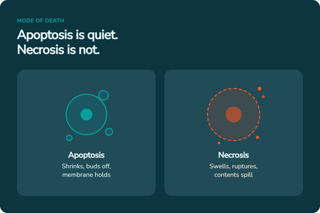 Application NoteWhy Are My Cells Dying? A Troubleshooting Guide to ApoptosisHow to tell apoptosis from necrosis, the six reasons cultures die, and how to confirm the mode of death with...
13 min read· Sep 2026
Application NoteWhy Are My Cells Dying? A Troubleshooting Guide to ApoptosisHow to tell apoptosis from necrosis, the six reasons cultures die, and how to confirm the mode of death with...
13 min read· Sep 2026
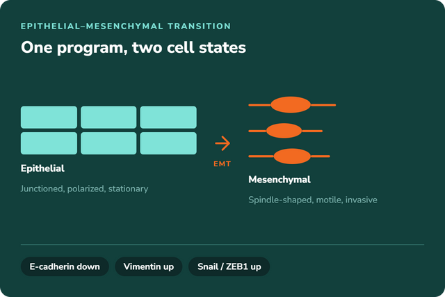 Application NoteEpithelial-Mesenchymal Transition (EMT) Explained: What It Does and When to Use ItThe EMT pathway, the epithelial cell markers and mesenchymal markers that define each state, the EMT transcription factors that drive...
12 min read· Sep 2026
Application NoteEpithelial-Mesenchymal Transition (EMT) Explained: What It Does and When to Use ItThe EMT pathway, the epithelial cell markers and mesenchymal markers that define each state, the EMT transcription factors that drive...
12 min read· Sep 2026
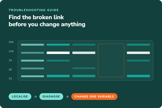 Why Your Western Blot Optimisation Experiment Isn't WorkingA four-step diagnostic that localises where a western blot broke, the common problems by symptom, and the single change to...
12 min read· Sep 2026
Why Your Western Blot Optimisation Experiment Isn't WorkingA four-step diagnostic that localises where a western blot broke, the common problems by symptom, and the single change to...
12 min read· Sep 2026
Recently viewed
Recently viewed products
Try Celltrypse Free – Request Your Sample Today
Experience the power of Celltrypse™, c-LEcta's innovative enzyme solution for gentle and efficient cell dissociation. Request your free sample and discover a superior alternative for your cell culture workflows.

Try Celltrypse Free – Request Your Sample Today
Experience the power of Celltrypse™, c-LEcta's innovative enzyme solution for gentle and efficient cell dissociation. Request your free sample and discover a superior alternative for your cell culture workflows.
